ETU – XImages
X Images is used for acquisition of X-ray maps collected by WDS and EDS spectrometers. The X-ray images can be collected simultaneously with a secondary or backscattered electron scanning electron microscope (SEM) image of the sample.
Selection of X-ray spectrometers and elements
The upper portion of the X Images panel is used to select the elements for mapping with WDS and EDS.
WDS
Tuning
Use the controls in the ETU ‘XLive’ panel to move the WD spectrometers to the elemental X-ray peaks of interest.
Choosing WDS elements for mapping
One element per WDS spectrometer may be included in the X-ray map acquisition. Press the Element/X-ray button to include individual WD spectrometers in the mapping.
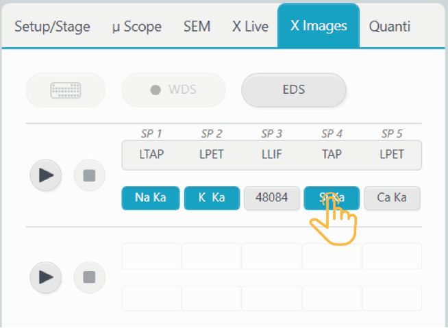
EDS
Choose the EDS spectrometer mode by pressing on the ‘EDS’ button  . Up to ten elements may be added to the EDS table. Individual X-ray maps will be acquired for all selected EDS lines. Selection of at least one element by EDS will activate acquisition of EDS hypermap data, which includes a full EDS spectrum at each pixel of the map. After map acquisition, a map of any element measured by EDS can be extracted from the hypermap data with the SX-Results program.
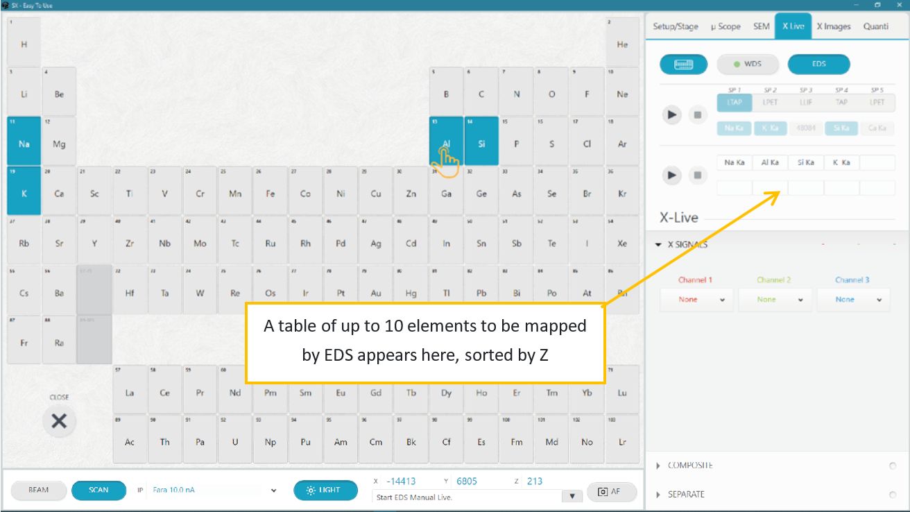
Press the ‘Periodic Table’ button  . Press on an element to add it to the EDS table. Press on the element a second time to remove it from the table.
X Images acquisition
The lower portion of the X Images panel is dedicated to image parameter setup and acquisition.
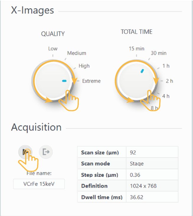
Two parameters can be user-adjusted: Quality and Total Time.
Quality affects the resolution of the images, from Low (coarse) to Extreme (fine).
Total Time is the estimated time required to acquire the images.
The remaining parameters are determined by Quality and Total Time, and by the current instrument configuration.
• Scan mode is determined by Scan Size. If Scan size <75 µm, the images are scanning the Beam. If Scan size is ≥ 75 µm, the images are made by scanning the Stage.
• Step size is calculated by Scan size/(# pixels in X axis)
• Definition is determined by Quality
• Dwell time (ms) is determined by the Quality (# of pixels) and Total Time.
Designate a filename for the image file, then press the ‘Run’ button
 to start an X Images acquisition.
The ongoing acquisition is visible in the SxSAB window. The acquisition completes after one frame.
Press the ‘Exit’ button
 to stop the acquisition.
File saving
The SX Images images are saved as ‘impDat’ files.
‘impDat’ files are proprietary image formats developed by CAMECA. They contain additional information, such as XYZ position and instrument configuration. These files can be viewed only with CAMECA software.
The files are saved in the Images&Profiles folder within the currently defined path (Project/Sample) and can be opened in the SX-Results program for additional image processing, including exporting to other file formats, such as TIFF, JPEG, and BMP.
Related Article
ETU – Introduction
Reading Duration 2min
Why a complex instrument should use a complex interface ? Trying to address this question leads us to rethink all of our softwares to turn them really easy to use. We had to forget all the way we develop softwares since decades and from a white paper, we imagined a new design that can hold advanced functionality together with intuitive touch-screen control in a unique window.
Start With Tactis ETU Software
Reading Duration 6min
Tactis ETU Software is a part of PeakSight 6.3 and late.
ETU – Setup/Stage
Reading Duration 4min
This part describes the SETUP/STAGE tab
Optical Microscopy
Reading Duration 4min
This article presents, in a tutorial mode, how to display and acquire high-quality images from optical microscopy system of the SX-FIVE. Functions are spread into ’Optical microscopy’ (all the hardware controls), ‘Save’ and ‘Mosaic’ subsections.
Scanning Electron Microscopy
Reading Duration 11min
Electronic images are provide though the Scanning Electron Microscopy functionalities of the SX-FIVE. Three detectors are available.
ETU – XLive
Reading Duration 10min
X Live is a powerful tool designed to draw real time X images using WDS or EDS detection. Images can be acquired separately or as a composite including SEM image as background.
ETU – Quanti/Check
Reading Duration 18min
The ‘Quanti’ tab provides all the tools necessary to get quantified chemical analysis of any solid sample, but also a range of check procedures to ensure the instrument hardware is well set. Both of them can be reach though the sub-tabs ‘Quanti’ and ‘Check’.
ETU – Configuration – Menu
Reading Duration 10min
Configuration window allows to set ‘Focus Auto Config’, ‘Astigmatism Auto Config’, ‘Expert System Config’, ‘Check standard Config’ and ‘Check Machine Config’.
Contact

Our support team is ready to help you
FAQ

Answers to some common questions from SX users
Troubleshooting

Quickly search for solutions to instrument issues